A digital database on the immunophenotypical cell profiles of the developing human brain is highly requested. The accumulation of data on human brain development, including data on the immunophenotype of various brain regions, remains an important task that has both fundamental and practical importance (for more information see "Modern trends in brain mapping and atlasing") .
Therefore, the Human Prenatal Brain Development Atlas (HBDA) has been launched, which aims to create spatiotemporal reference maps of the whole foetal brain. It will combine histological and immunohistochemical methods. The HBDA could help answer questions in the histological brain structure, developmental biology, and cell fate and lineage, including neuro- and gliogenesis.
Tissue sources and processing.
The HBDA is based on postmortem human brain autopsies from the unique #Collection of the Prenatal Human Development of the Laboratory of the Nervous System Development of the Research Institute of Human Morphology. It consists of the material from the second post conceptional week (2 pcw) until birth. The materials have been collected in the last five decades and are currently being renewed. The collection consists of more than 200 formalin-fixed human embryos and foetuses at different gestational stages. The collection contains not only fixed material but also histological, immunohistochemical, and electron-microscopic preparations. All the material was taken and handled according to national legislation and the Declaration of Helsinki. All protocols were approved by the local Ethics Committee of the Research Institute of Human Morphology.
The classification of the foetal period, in which it is divided into four stages (pre-foetal (10–12 gestational weeks (gw)), early foetal (13–20 weeks), middle foetal (21–28 weeks), and late foetal (29–40 weeks) periods)) is used (Milovanov and Saveliev, 2006).
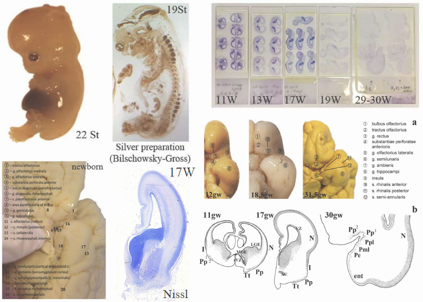
Histological preparation
Most samples are buffered formalin-fixed. For the prenatal reference Atlas, serial coronal and sagittal 5–10 μm thick paraffin sections from the whole embryo or foetal head, foetal brain, or an entire hemisphere, depending on the developmental stage, are stained with hematoxylin and eosin, Mallory trichrome, and Nissl techniques.
Immunohistochemistry (ICH)
A combination of the classic cytoarchitecture with recent immunohistochemical methods is a great advantage of the immunomorphological approach that resulted in the regional-specific immunophenotyping of certain brain areas at various prenatal stages. In accordance with the HBDA objectives, immunohistochemical experiments are planned using direct immunoperoxidase, double, and multiplex labeling with immunofluorescence and chromogen methods directly on a series of histological sections, followed by an analysis of the results using morphometry.. Multiplex immunolabeling is used for the spatial analysis of antigen patterns on the same slice. Thus, immunophenotypes of the whole brain structure are obtained as a result of the procedure.
The mechanisms and factors of the differentiation between multipotent and oligopotent progenitor cells of the nervous system are central problems in recent developmental neurobiology. The choice of a marker panel for the project is based not only on the authors’ own experience but also on the published data and online materials on the related sources such as The Human Protein Atlas, Allen Brain Atlas, and others. Preference in each phase of experiments is given to markers that are most well-established in the world research and medical practice and consistently work on human foetal autopsy material. Thus, the HBDA will be useful for the comparative analysis with other research groups’ data
Human Brain Imaging
Annotated atlases based on the series of virtual slices of sixteen human foetal brains in different developmental stages will be a main component of the HBDA.
The basic annotated atlases will contain an image library of virtual sections with a zoom tool corresponding to an objective resolution of up to ×20. Virtual slices from the serial histological preparations of human foetal brain autopsies will be made using the MECOS-C2 system, specially modified for large (non-standard) preparation sizes (up to 12×9 cm). The system collects hundreds of images in lengthwise strips, which are later stitched together to create one large image. The exposure time, white balance, and flat-field correction are set independently for each slide. The acquired SVS files subsequently will be converted to JPEG. As the initial step of the project, histological preparations from the collection will be digitized, annotated, and analysed to create a reference base for the Atlas. New preparations will be prepared especially for the HBDA .
Data organization. Neuroanatomical parcellation. Annotation of sections
A separate task is marking and annotating virtual preparations for each developmental stage. Annotation schemes will be overlapped on the digital images of histological slides. The resulting vector graphics will be converted to Scalable Vector Graphics (SVG). Each vector area will then be associated with a certain brain structure and annotated; the collating vector in this way allows the flexibility to create various presentation modes (e.g., with or without colorization and transparency).
There is no one uniform nomenclature for all structures of human developing telencephalon. Structural delineations will be based on classic adult human brain morphology and special publications on the certain structure developmental course with special references for evolutionary nomenclature principles. All published sources used for each structural complex will be provided as special comments. Differences between existing neurologic traditions will be compared and commented on, with special references to classic German, American, and Russian (Soviet Union) ontological principles and the recent "standard" of anatomical ontology provided in Nieuwenhuys et al. (2008), Terminologia Neuroanatomica (Ten Donkelaar et al., 2017) and the Allen Brain Atlas as an online source (https://atlas.brain-map.org/). The structure will be delineated and annotated following comparative evolutionary and ontogenetic criteria – from the general to the particular, from complex structures to parts and subdivisions according to the developmental course. The preliminary marking is done according to the Allen Brain Atlas as an online source (https://atlas.brain-map.org /)
References:
- Milovanov AP, Saveliev SV (2006) Rational periodization and methodical aspects of embryology. In: Milovanov AP, Saveliev SV (eds) Prenatal human development. MDV, Moscow, pp 21–32 Russian
More information about modern trends in brain mapping and atlasing